
Pictorial supplement to The Fifth Kingdom - Chapter 11a
Fungal Ecology - part 1
(41 pictures)The Fungal Succession on Herbivore Dung
Zygomycetes
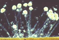
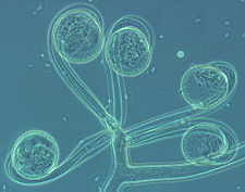
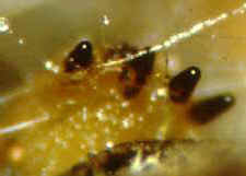
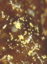
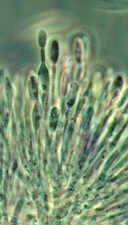
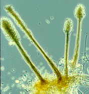
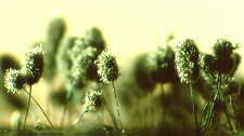
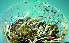
Coprophilous fungi - Pilobolus
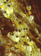
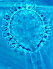
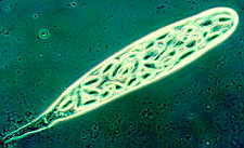
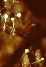
Coprophilous fungi - Pilobolus sporangiophores and sporangia.
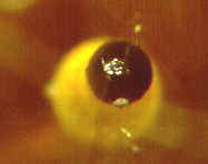
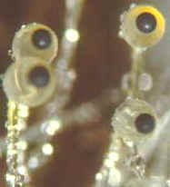
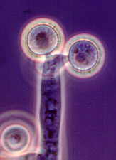
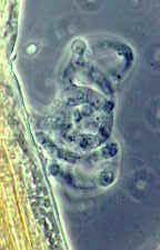
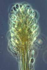
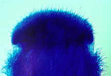
Coprophilous fungi - Pilobolus - a single sporangium and its subsporangial vesicle. The yellow colour of the carotenoid retina is refracted in the vesicle.
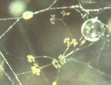
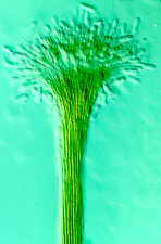
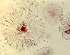
Coprophilous fungi - branched sporangiophores of Piptocephalis.
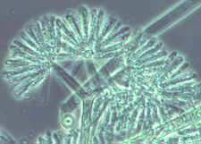
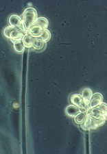
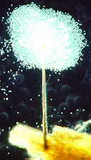
Coprophilous fungi - Piptocephalis heads, showing clustered fans of merosporangia.
Coprophilous fungi - sporangiophores of Syncephalis (low power).
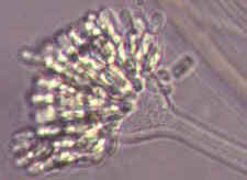
Coprophilous fungi - one sporangiophore head of Syncephalis, showing radiating merosporangia (high power).
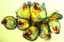
Coprophilous fungi - Circinella minor (1) - young recurved sporangia
Coprophilous fungi - Circinella minor (2) - sporangia expanding
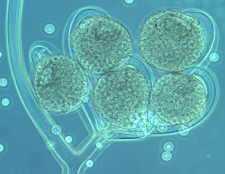
Coprophilous fungi - Circinella minor (3) - mature columellate sporangia
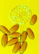
Coprophilous fungi - Rhopalomyces elegans - apical vesicle with unispored sporangia.
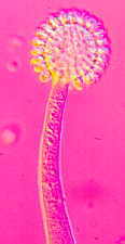
Coprophilous fungi - Cunninghamella - apical vesicle and unispored sporangia. Ascomycetes
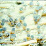
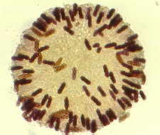
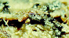
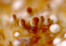
Coprophilous fungi - Ascobolus - numerous tiny apothecial ascomata
Coprophilous fungi - Ascobolus - several ascomata
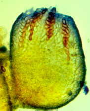
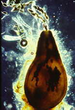
Coprophilous fungi - Ascobolus - single ascoma. Note coloured ascospores and protruding ascal tips.
Coprophilous fungi - Ascobolus - protruding, phototropic ascal tips.
Coprophilous fungi - Saccobolus - a squash of an entire apothecial ascoma. The dark bodies are the adherent octets of ascospores (see below)
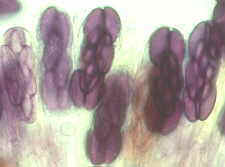
Coprophilous fungi - Saccobolus - some of the asci more highly magnified, showing that the 8 purple spores stick together and are discharged as a unit.
Coprophilous fungi - many-spored asci of Thecotheus
Coprophilous fungi - one ascus of Thecotheus. How many spores?
(clue - it must be a multiple of 8)
Coprophilous fungi - black perithecial ascomata of Podospora - look closely and you will see several phototropic necks, pointing to the right.
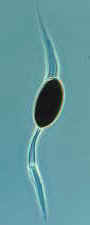
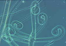
Coprophilous fungi - Podospora - a perithecial ascoma, and a single ascospore with one primary (tubular) and two secondary (gelatinous) appendages. Hyphomycetes
Coprophilous fungi - Basifimbria - on the substrate (left) and under high power (right) - note the sympodial conidium development
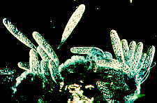
Coprophilous fungi - tall conidiophores of Arthrobotrys on natural substrate.
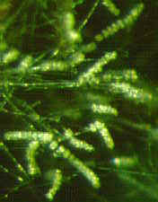
Coprophilous fungi - clavate didymospores of Arthrobotrys developing in synchronous clusters.
Coprophilous fungi - a hyphal network of Arthrobotrys which functions as a nematode trap.
Coprophilous fungi - Graphium synnematal conidiomata on the substrate, each with an apical drop of slimy spores (left), and one conidioma mounted on a slide (right). The vast numbers of spores obscure the structures that produce them (but see below)
Coprophilous fungi - a synnematal conidioma of Graphium (left) and details of the percurrently extending conidiogenous cells under oil immersion and phase contrast (right).
Coprophilous fungi - synnematal conidiomata of the dry-spored Cephalotrichum.
Coprophilous fungi - a synnematal head of Cephalotrichum, with percurrently extending conidiogenous cells.
Coprophilous fungi - Trichurus, another coprophilous synnematal hyphomycete with twisted, hair-like setae arising all over the fertile head. Basidiomycetes
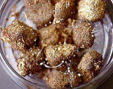
Coprophilous fungi - primordia of Coprinus developing on horse dung.
Coprophilous fungi - close-up of the Coprinus primordia.
Coprophilous fungi - a stained and mounted primordium of Coprinus.
Coprophilous fungi - profuse fruiting of Coprinus on horse dung.
Coprophilous fungi - delicate caps of Coprinus stuck to the glass lid of the incubation dish.
Go to
Chapter 11b
Go to Table of Contents
© Mycologue publications 2020