
Pictorial Supplement to The Fifth Kingdom - Chapter 17
Mycorrhizae - mutualistic plant-fungus symbioses
(35 pictures)[grateful acknowledgment to S. Berch, C. Godbout, M. Brundrett,
D. Malloch & J.M. Trappe who generously made some of these
images available to me for teaching purposes]

dichotomously branched ectomycorrhizas of a basidiomycete with a conifer.
X 4

ectomycorrhizas of Suillus subluteus with Pinus resinosa (the branched ends of the short lateral roots)
X 2/3

dichotomous ectomycorrhizas (upper) and mycelial strands (lower) of Amanita muscaria on Pinus strobus.

ectomycorrhizas of Laccaria bicolor with Populus tremuloides.
X 3

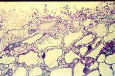
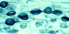
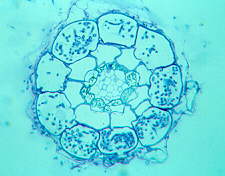
transverse section of an ectomycorrhiza of Pseudotsuga menziesii with Rhizopogon colossus showing the fungal mantle (brown in this example).
X 50
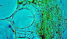
section of outer layers of an ectomycorrhizal root of Pinus strobus, showing some of the mantle and the Hartig net - the latter formed by hyphae of the mycobiont, Pisolithus tinctorius, penetrating between the cortical cells of the root.

Ectomycorrhizas - a root cell completely surrounded by hyphae of the Hartig net.

mantle of an ectomycorrhiza of Populus tremuloides in section and Hartig net in surface view - the section was cleared then stained with chlorazol black E. The fungus is stained dark brown. Viewed with interference contrast. (see Fig 52 in Brundrett et al. 1990. Can. J. Bot. 68: 551).

surface view of the uniquely branched and contorted hyphae of the Hartig net filling the space between cells of the root cortex of an ectomycorrhiza.
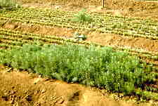
seedlings of Douglas fir with and without ectomycorrhizal partners.

bisected basidioma of Pisolithus tinctorius, an important ectomycorrhizal fungus, showing the gleba divided up into locules, and the yellow pigment in the base.
(2) Endomycorrhizas (Arbuscular Mycorrhizas)

a part of the "extramatrical" mycelium of Glomus mosseae, an endomycorrhizal fungus grown in leek roots in a root chamber. The sand around the roots has been carefully rinsed away, and the material cleared then stained with chlorazol black E (M. Brundrett)

as above, but showing monosporic sporocarps of Glomus mosseae with hyphal peridium. (M. Brundrett)
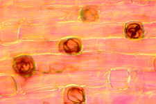
long and short cells in the dimorphic exodermis of Smilacina racemosa. The short cells remain unsuberized longer, and permit entry of endomycorrhizal fungi. Five of the 8 short cells shown here have been colonized. (see Fig 39 in Brundrett & Kendrick 1990 New Phytol. 114: 469)

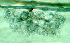
penetration pattern of Glomus versiforme, an endomycorrhizal fungus, into a leek root. Note that arbuscules are formed in cells of the cortex, and that the fungus does not penetrate the stele. (see Fig 20 in Brundrett et al. 1985 Can. J. Bot. 63: 184)
a stained arbuscule of Glomus mosseae in a leek root cell (a superb photomicrograph by Mark Brundrett - see Fig 17 in Brundrett et al. 1984 Can. J. Bot. 62: 2128)

hyphae and arbuscules of an endomycorrhizal fungus in Asarum (wild ginger) (see Fig 15 in Brundrett & Kendrick 1988 Can. J. Bot. 66: 1153)

colonization of a root by an endomycorrhizal fungus. Note hyphae, arbuscules and vesicles. (see Fig 21 in Brundrett et al. 1985 Can. J. Bot 63: 184)
a later stage of colonization. The arbuscules contract and degenerate, and more vesicles develop.

a leek root packed with vesicles of its endomycorrhizal fungal partner.

these structures in the "roots:" of early land plants fossilized in the Rhynie Chert (350 MYBP) are regarded as vesicles of an early endomycorrhizal fungus.

endomycorrhizas - extramatrical spores of Glomus versiforme (M. Brundrett).

endomycorrhizas - a single extramatrical spore of Glomus mosseae (M. Brundrett)
section of a sporocarp of Glomus, an endomycorrhizal fungus.

the "big plant - little plant" experiment showing the benefits of the endomycorrhizal symbiosis (plants on the right are not mycorrhizal).
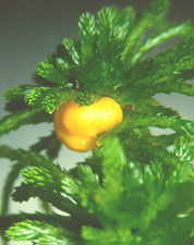
Endogone pisiformis fruiting on Sphagnum in a bog. The yellow sporocarp contains a very large number of zygosporangia (below). Though formerly recognized as the type genus of endomycorrhizal fungi, Endogone is now known to be ectomycorrhizal.

a single zygosporangium of Endogone pisiformis. Note the two suspensors,
(3) Arbutoid mycorrhizas (Ectendomycorrhizas)
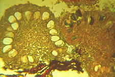
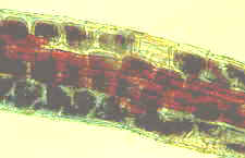
Arbutoid mycorrhizas of Arbutus menziesii with Lactarius deliciosus.

Transverse section of arbutoid mycorrhiza of Arbutus menziesii with Lactarius deliciosus. Note mantle, and cortical cells filled with hyphae.

Arbutoid mycorrhiza - high power view of part of section shown above.
(4) Ericoid mycorrhizas
ericoid mycorrhiza of salal, Gaultheria shallon. Dark blobs are masses of fungal hyphae in cortical cells of root.
transverse section of ericoid mycorrhiza of salal, Gaultheria shallon.
(5) Monotropoid mycorrhizas
section of part of a monotropoid mycorrhiza of Monotropa uniflora. Note the diagnostic peg-like hypha penetrating a root cell (indicated by arrowhead).
(6) Orchid mycorrhizas

root cells of Goodyera oblongifolia containing coils (pelotons) of hyphae (probably of a mycorrhizal Rhizoctonia).

Goodyera cells again: note two distinct constrictions in the fungal hyphae where they penetrate from one cell to the next. For more information on mycorrhizas and more pictures, visit this site:
https://mycorrhizae.com/
Go to Chapter 18 Go to Table of Contents
© Mycologue publications 2020