
Pictorial supplement to The Fifth Kingdom - Chapter 11c
Fungal Ecology - part 3
Fungal succession on needles of Scots pine (Pinus sylvestris)(23 pictures)
This story is derived from my Ph.D. work of many years ago...BK
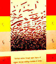
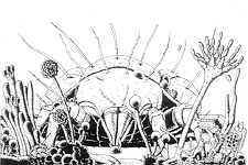
Vertical section through the organic horizon of a pine forest, showing changing orientation of fallen needles with time, and their transition from L > F1 > F2 > H layers, which takes 9 years.

the five categories of needles I recognized. From left to right - (1) living and green (on the tree), (2) dead, pale brown, tough (L layer), (3) darker but still tough (F1 layer), (4) blackish and softer (F1), (5) greyish and fragmenting (F2).

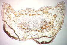
transverse section of a healthy pine needle - this is how things start out... Lophodermium may well be living inside the needle, but is asymptomatic. Aureobasidium (Pullularia)will almost certainly be present on the surface, but as an inconspicuous saprobe.

when needles die and fall, entering the L layer, the Lophodermium soon fruits, producing covered, lenticular ascomata (see next picture)...
transverse section of a dead needle from the L layer, passing through two Lophodermium ascomata which have developed just beneath the epidermis.

section of another needle from the L layer, this one colonized by Fusicoccum, which has produced two pycnidial conidiomata.

the surface of needles in the F1 layer are almost covered by a network of dark fungal hyphae, which give rise to a variety of conidiophores...
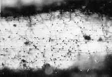
...such as those of Slimacomyces (formerly Helicoma), whose dark conidia can be seen here...

Slimacomyces conidiophores and single, non-deciduous helicospores.
...or those of Sympodiella (a new genus)...
a Sympodiella conidiophore, showing its unique combination of sympodial extension and the production of thallic-arthric conidia.

...or sometimes sclerotia and conidiophores of Thysanophora (another new genus).

partial conidiophores of Thysanophora penicillioides.

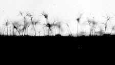
...or the tall conidiophores of Verticicladium (the anamorph of the discomycete, Desmazierella).
Note that most of these occur in pure stands. This is because needles tend to become dominated by one or two species...

This needle is partitioned between two very different-looking fungi, and the dividing line is often very sharp, as you can also see below...

Here, the needle section shows that there is often a sharp melanin-coated line of demarcation between neighbouring fungal fiefdoms.
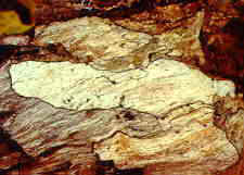
Exactly the same thing often happens in decaying wood, on a much larger scale. Such wood is often described as "spalted."
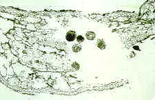
Now, in the F2 layer, the fungi have done their job, and the animals can come in and eat the "conditioned" or predigested needle tissue. Here a beetle mite (see below) has eaten its way through the interior of the needle, leaving its faeces to tell the story.
beetle mites (Acari) are heavily armoured fungus- and needle-eaters
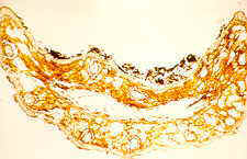
Now the interior has been eaten out, and the surface of the F2 layer needle is often encrusted with faecal deposits which contain many spores of the fungi illustrated above.

The over-all picture, following the pine needles through 9 years of their mainly fungal decay.

compare the biomass of other organisms in forest soil with that of the fungi...
References on needle decay:
Kendrick, B. 1958. Microfungi in pine litter. Nature 181: 432.
Kendrick, B. 1958. Helicoma monospora sp. nov. from pine litter.
Trans. Brit. mycol. Soc. 41: 446-448. [later made the type species of Slimacomyces Minter]
Kendrick, B. 1958. Sympodiella, a new hyphomycete genus.
Trans. Brit. mycol. Soc. 41: 519-521.
Kendrick, B. 1959. The time factor in the decomposition of conifer[ous] leaf litter.
Can. J. Bot. 37: 907-912.
Kendrick, B. 1961. Hyphomycetes of conifer leaf litter. Thysanophora gen. nov.
Can. J. Bot. 39: 817-832.
Kendrick, B. and A. Burges 1962. Biological aspects of the decay of Pinus sylvestris leaf litter. Nova Hedwigia 4: 313-342.
Go to Chapter 12 Go to Table of Contents
© Mycologue publications 2020